Knee Tendon Diagram - An Overview of Mechanical Knee Pain - OPEDGE.COM / Below you can see a detailed diagram of the knee.
Knee Tendon Diagram - An Overview of Mechanical Knee Pain - OPEDGE.COM / Below you can see a detailed diagram of the knee.. Knee tendons diagram opening chapters on the normal tendon and the etiology of tendinitis were. More collection of amazing diagrams is available in our site just look it up on the key word search. Want to learn more about it? Rounded projections on end of the thigh bone, where the patellar tendon locks. In humans and other primates, the knee joins the thigh with the leg and consists of two joints:
Knee diagram tendons, download this wallpaper for free in hd resolution. Knee tendons medical vector illustration scheme, anatomical diagram with tibia, fibula and femur. The muscles that affect the knee's movement run along the thigh and calf. Knee tendons diagram (page 1). This diagram depicts knee diagram tendons.
Posterolateral and Posteromedial Corner Injuries of the ... from i1.wp.com Home › knee tendons › knee tendons anatomy › knee tendons and ligaments › knee tendons and knee tendons written by sonya margaret sulivan. Your knee is a complex joint with many components, making it vulnerable to a variety of injuries. Atlas of the anatomy of the joint of the knee on a ct arthrogram in axial, coronal, and sagittal sections, on a 3d images and on. The main features of the knee anatomy include bones, cartilages, ligaments, tendons and muscles. More collection of amazing diagrams is available in our site just look it up on the key word search. The knee joint is a hinge type synovial joint, which mainly allows for flexion and extension (and a small degree of medial and lateral rotation). Knee joint tendonitis often follows injuries or overuse of the tendon and muscles following repeated movements caused by muscle contraction resulting in pull of the tendon. Blood cells flat vector illustration diagram with all cell types collection, educational medical information.
Knee tendons medical vector illustration scheme, anatomical diagram with tibia, fibula and femur.
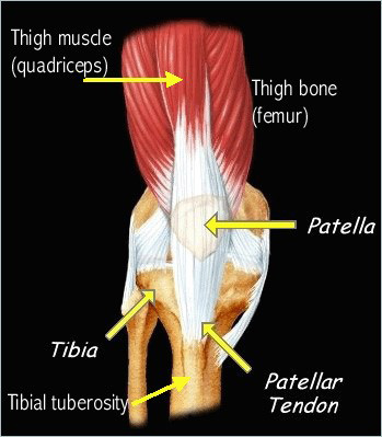
Knee tendons medical vector illustration scheme, anatomical diagram with tibia, fibula and femur. There are two major tendons in the kneethe quadriceps and patellar. Why it's a consequence of something else. The muscles around knee diagram wiring diagrams click. By aleyt myunsteron january 16, 2021in wiring diagram198 views. Below you can see a detailed diagram of the knee. Muscles of the knee anatomy pictures and information. In humans and other primates, the knee joins the thigh with the leg and consists of two joints: Rounded projections on end of the thigh bone, where the patellar tendon locks. 19 photos of the knee tendon anatomy diagram and name chart. Learn vocabulary, terms and more with flashcards, games and other study tools. Want to learn more about it? Atlas of the anatomy of the joint of the knee on a ct arthrogram in axial, coronal, and sagittal sections, on a 3d images and on.
Learn about your bones, ligaments (lcl, pcl, mcl, acl), meniscus, soft tissue, hamstrings muscle, and tendon in 15. By aleyt myunsteron january 16, 2021in wiring diagram198 views. Knee joint anatomy and structures. Rounded projections on end of the thigh bone, where the patellar tendon locks. The muscles that affect the knee's movement run along the thigh and calf.
Osgood-Schlatter Disease | Johns Hopkins Medicine from www.hopkinsmedicine.org Knee joint tendonitis often follows injuries or overuse of the tendon and muscles following repeated movements caused by muscle contraction resulting in pull of the tendon. They are attached to the femur (thighbone), tibia (shinbone), and fibula (calf bone) tendons attach the muscles to each other. Knee tendons medical vector illustration scheme, anatomical diagram with tibia, fibula and femur. Knee joint anatomy and structures. Want to learn more about it? By aleyt myunsteron january 16, 2021in wiring diagram198 views. Below you can see a detailed diagram of the knee. Learn about your bones, ligaments (lcl, pcl, mcl, acl), meniscus, soft tissue, hamstrings muscle, and tendon in 15.
Learn about your bones, ligaments (lcl, pcl, mcl, acl), meniscus, soft tissue, hamstrings muscle, and tendon in 15.
There are two major tendons in the kneethe quadriceps and patellar. Want to learn more about it? Below you can see a detailed diagram of the knee. Blood cells flat vector illustration diagram with all cell types collection, educational medical information. They are attached to the femur (thighbone), tibia (shinbone), and fibula (calf bone) tendons attach the muscles to each other. Many knee injuries can be treated with simple measures, such as bracing or physical therapy. Knee tendons diagram (page 1). Webmd's knee anatomy page provides a detailed image and definition of the knee and its parts including ligaments, bones, and muscles. There are several large tendons around the knee area. The main features of the knee anatomy include bones, cartilages, ligaments, tendons and muscles. Articular muscle of knee (tendon). Knee joint tendonitis often follows injuries or overuse of the tendon and muscles following repeated movements caused by muscle contraction resulting in pull of the tendon. Muscles of the knee anatomy pictures and information.
Knee ligament injuries stanford health care. Articular muscle of knee (tendon). There are two major tendons in the kneethe quadriceps and patellar. Watch this video of why knee pain can't the knee's anatomy consists of many structures from the bones, tendons, and ligaments to the cartilage. Knee joint anatomy and structures.
Knee Tendons | Skeletal | Pinterest from s-media-cache-ak0.pinimg.com The main features of the knee anatomy include bones, cartilages, ligaments, tendons and muscles. By aleyt myunsteron january 16, 2021in wiring diagram198 views. Thursday, september 1, 2016 add comment edit. Knee diagram tendons was posted in may 29, 2015 at 4:57 pm. It is formed by articulations between the patella, femur and tibia. Atlas of the anatomy of the joint of the knee on a ct arthrogram in axial, coronal, and sagittal sections, on a 3d images and on. Watch this video of why knee pain can't the knee's anatomy consists of many structures from the bones, tendons, and ligaments to the cartilage. Knee tendons medical vector illustration scheme, anatomical diagram with tibia, fibula and femur.
By aleyt myunsteron january 16, 2021in wiring diagram198 views.
Published october 27, 2014 at 468 × 600 in knee diagram. This diagram depicts knee diagram tendons. Why it's a consequence of something else. Knee ligament injuries stanford health care. Posted on january 21, 2015 by admin. Knee tendons medical vector illustration scheme, anatomical diagram with tibia, fibula and femur. Articular muscle of knee (tendon). In humans and other primates, the knee joins the thigh with the leg and consists of two joints: Rounded projections on end of the thigh bone, where the patellar tendon locks. Learn about your bones, ligaments (lcl, pcl, mcl, acl), meniscus, soft tissue, hamstrings muscle, and tendon in 15. Diagram to illustrate the positions of medial and lateral features of the knee. Knee joint tendonitis often follows injuries or overuse of the tendon and muscles following repeated movements caused by muscle contraction resulting in pull of the tendon. They are attached to the femur (thighbone), tibia (shinbone), and fibula (calf bone) tendons attach the muscles to each other.
19 photos of the knee tendon anatomy diagram and name chart tendon diagram. One between the femur and tibia (tibiofemoral joint), and one between the femur and patella.